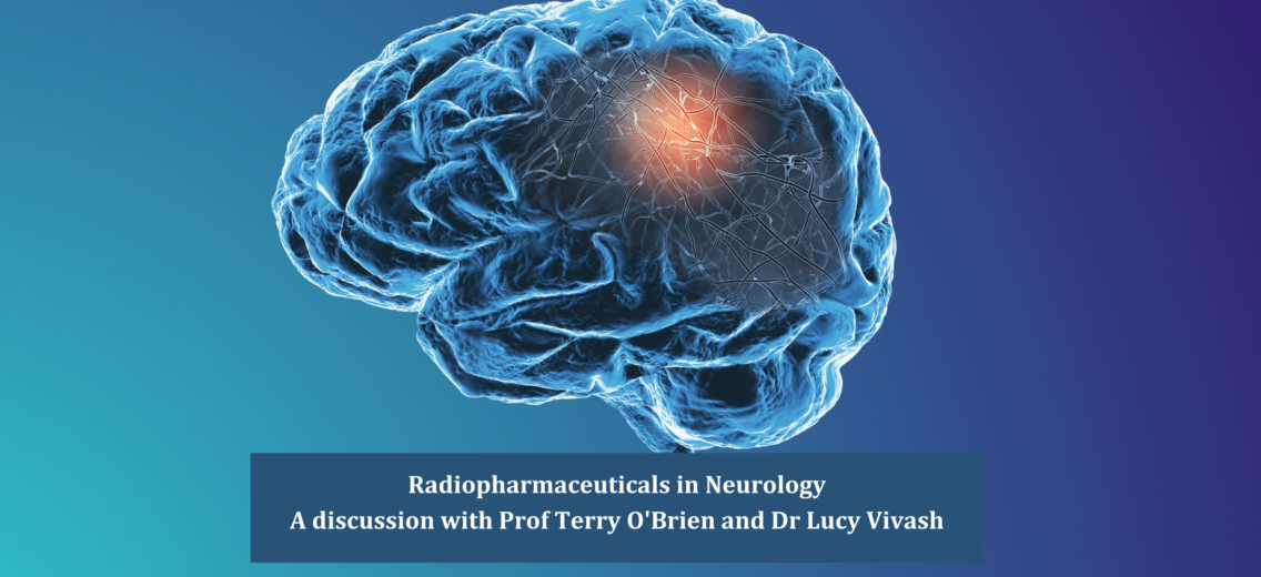
Cyclotek’s Media Associate, Natasha Jose Kalathiveetil sat down with Terry O’Brien and Lucy Vivash to discuss their collaboration with Cyclotek and the Monash Institute of Pharmaceutical Sciences to successfully complete their research endeavours
Professor Terence (Terry) O’Brien is Chair of Medicine (Neurology) and Head of The Central Clinical School at Monash University, and Program Director Alfred Brain and Deputy Director of Research at Alfred Health. Dr Lucy Vivash is a Research Fellow in the Department of Neuroscience at Monash University. Her research focuses on the application of neuroimaging (particularly PET) to better understand neurological and neurodegenerative diseases.
Can you tell us about the importance of PET brain imaging in patient diagnosis? How can better diagnosis improve patient outcomes?
Terry O’Brien – Brain imaging with PET is what we call a functional form of imaging, meaning that it shows us how the brain is functioning, as opposed to its structure like how we see with MRI scans.
PET imaging allows us to much more specifically understand the underlying pathological processes that are causing neurological diseases in patients. Diseases such as epilepsy or dementia may present as very similar in the symptoms that a patient experiences, but there are actually many different diseases that underlie these conditions. Neither epilepsy nor dementia are a single disease in themselves, rather they are a collection of symptoms derived from a whole variety of different underlying diseases.
The problem used to be that with just clinical assessment and the standard tools we had available, it was very difficult to distinguish one cause of disease from another. As medical science has advanced, we now have many therapies in development that are actually very specific for different pathological processes. However, in order to apply these therapies effectively, we need to understand and diagnose the underlying pathology of a patient presenting with a neurodegenerative condition. PET has allowed us to make those diagnoses and enables what we call precision medicine, where instead of a one-size-fits-all treatment, we are targeting treatment specifically to the cause of the disease in that particular patient.
Lucy Vivash – And it’s this use of neuroimaging broadly but especially using PET in differential diagnosis for patients early on, that could impact so many lives. Often patients can present with symptoms but cannot get a specific diagnosis until 3 years down the track, the disease has progressed so much that we can see it more clearly. With developments in PET, if we can have a neuroimaging technique that can indicate the pathology earlier on, it’s a lot better for counselling of the patient and even potential therapies.
Terry, you briefly mentioned the difference between PET and MRI’s, how does a brain scan look different in these two methods of imaging?
Terry O’Brien– PET and MRI are both important and very complimentary brain imaging investigations. An MRI is primarily structural imaging whilst PET is functional imaging. I describe structural imaging as like looking at a map, you see clearly where the roads and houses are, but with PET imaging, you are able to see the cars moving on the roads, so where the function within that structure is occurring. Moreover, because we can have a number of different tracers that we can radiolabel using PET imaging, we can also see very specific neurological processes in a way that we can’t with MRI.
Lucy, how has brain imaging developed in the last decade or since you started working in research?
Lucy Vivash – PET in neurology has really come leaps and bounds in the last 10 years, there are so many more tracers that are high quality available now. For example, about 10 years ago we had our first amyloid tracer which really wasn’t very good and was limited to only very advanced research settings – now we have so many new and fantastic amyloid tracers that have become more widely accessible.
This recent explosion of PET has meant that we now have access to many new tracers that are fit-for-purpose and able to detect subtlety in the brains’ systems. Neurology has now switched on to what PET can actually do and how it can look at any protein within a system or target any specific pathology, and with the right tracer, you can actually see it.
What in your opinion was the cause of this recent explosion of interest in PET in the neurology space?
Lucy Vivash – With the developments in cancer biology and therapeutics, people were all of a sudden investing in PET for molecular targets in cancer. We saw how this precision medicine had enabled all these new treatments in oncology, so we thought, let’s give PET another shot in neurology!
The advances in our understanding of the pathophysiology of neurological diseases has resulted in this perfect storm, which has enabled the application and development of PET. What’s really exciting for me about these technological advances is that PET is now becoming available to so many more people.
Terry O’Brien – I absolutely agree with Lucy that neurology has very much been led by the oncology field. 30 years ago, in cancer therapeutics we used the same chemotherapy for a broad range of tumours, now we molecularly type the tumour and provide therapies that are focused on the molecular mechanism of the cancer. This has made an enormous difference to the outcomes of many oncology patients that were previously very difficult to treat.
Similarly, not too long ago in neurology, we were still using a one-size-fits-all treatment approach and for many diseases, we actually had no effective therapies. As we’ve come to understand more about the molecular basis of many neurological diseases, we have adopted that same thinking as in oncology, where focus on targeting the underlying processes. The tricky thing with neurology is that you need to create non-invasive ways of typing the pathology and that is where PET has really stepped in and allowed us to image the pathology in a living human, which is essential to precision medicine.
Can you tell us a little bit more about Cyclotek’s 18F Florbetaben? What do you expect to see on a PET scan using amyloid agents?
Lucy Vivash – Florbetaben is a newer amyloid tracer, essentially what it does is it binds to the amyloid proteins in the brain, which are associated with Alzheimer’s disease. In a typical amyloid PET scan, if you don’t have Alzheimer’s, the amyloid tracer will bind to the white matter of the brain, however, if you have a lot of amyloid, we see the tracer bind to the cortical area of the brain in a different pattern. Not everyone with high amounts of amyloid protein will have Alzheimer’s disease, but almost all Alzheimer’s disease patients will present with high amyloid proteins. Hopefully, in the near future, the TGA will get around to making some of these tracers available outside of research for wider use.
Terry O’Brien – Absolutely, we’re just emerging on the phase where treatments for Alzheimer’s disease are going to enter practice. In the last decade, there has been a lot of clinical trials and PET scans have been essential in ensuring that the right patients are enrolled in those trials. This year we’ve just had the first treatment approved by the FDA for a disease-modifying treatment for Alzheimer’s disease, and it may well be approved by the TGA in the near future. Because these drugs are very expensive, we expect it will almost certainly be a prerequisite that patients have a positive amyloid PET diagnosis to gain access to this clinical treatment.
Moving onto Life Molecular Imaging’s 18F PI-2620 compound targeting TAU, can you tell us about your research here?
Lucy Vivash – TAU is relevant to almost every neurodegenerative disease that exists, but the disease we’re really focused on for TAU diagnosis and therapy is frontotemporal dementia, whereby about 45% of patients have a TAU based pathology.
The 18F PI-2620 tracer is really exciting. I remember talking to Cyclotek’s CEO Greg Santamaria about this 5 years ago, he was showing me the latest in TAU tracers and out of a few TAU tracers we selected for research, we chose PI-2620. In an optimistic fashion, Terry and I wrote hundreds of grant applications for research looking at new treatments that are anti-TAU for some of these neurodegenerative diseases.
Eventually, we got the funding as well as the phone call that PI-2620 was available. Having a look at some of the first data of the PET scan was a really memorable moment for me; you could see the TAU was right where it should be, mapping perfectly with the severity of the disease in these patients! Now we’re using PI-2620in our clinical trials. Theoretically, this is the future, because if our drug works the way we think it does and reduces TAU in the brain, then we will see on the PET scan an absolute definitive change in TAU levels prior to and after treatment.
What role does PET imaging play in the future of neurology? How would you expand its use or explore its potential?
Terry O’Brien – Once we develop new treatments that are effective in slowing down or reversing a neurological disease, we need ways to monitor the effectiveness of these treatments. Potentially, PET will be a way of seeing if we have effectively addressed the pathology as we’d wanted to.
The other area that we’re expecting to become popular soon in the neuroscience area is theranostics. Theranostics combines the diagnostics with therapies by attaching some sort of therapy to your targeted tracer. The tracer which binds to a particular type of protein or cell carries with it something which either kills or modulates the protein or cell, which will make for very targeted and personalised therapy.
I expect it will become part of the diagnostic algorithm, we won’t just diagnose someone with dementia or Alzheimer’s, but rather with Alzheimer’s disease-related to a particular type of amyloid pathology and therefore we can put them into a treatment program that targets that type of pathology. This is a whole new area of research we’re going to be getting into in neurology and it’s probably where the next 10 years is taking us.
Are you optimistic about the future of diagnosing and treating neurological disease?
Terry O’Brien – Absolutely! The last decade has really taken years of neuroscience research to the point where now therapies that target specific pathologies are coming into trials. For so many different diseases that we could never imagine would ever be treatable, there are now therapies being trialled.
Lucy Vivash – Like Terry, I’m extremely optimistic about the future of neurology. The developments that have come through in the last decade have really changed things and everyone is now taking in a wider approach and realising that we need to look at multiple different aspects of a disease and use multiple different techniques and technologies to do so. I think the future development and application of therapies will be closely intertwined with the research of new diagnostic tests, particularly with PET.